
Journal of Gastroenterology Research and Practice
Short Report - Open Access, Volume 5
Dysphagia caused by anterior cervical osteophytes: An interesting case report
Vimalraj V, MD1*; Premkumar K, DM2; Ratnakar Kini P, DM2
1Senior Resident, Institute of Medical Gastroenterology, Madras Medical College, Chennai, India.
2Professor, Institute of Medical Gastroenterology, Madras Medical College, Chennai, India.
*Corresponding Author : V Vimalraj
Senior Resident, Institute of Medical Gastroenterology, Madras Medical College, Chennai, India.
Tel: +91 8610258630;
Email: drvimaltnj@gmail.com
Received : Jun 04, 2025
Accepted : Jul 09, 2025
Published : Jul 16, 2025
Archived : www.jjgastro.com
Copyright : © Vimalraj V (2025).
Abstract
Dysphagia is a common symptom in the elderly, often resulting from mechanical or neuromuscular etiology. Cervical osteophytes are a rare but notable mechanical cause of dysphagia due to external compression of the esophagus. We report the case of a 61-year-old woman who presented with gradually progressive dysphagia over two years. Imaging with barium swallow, CT, and MRI revealed large anterior cervical osteophytes causing esophageal indentation. She was managed conservatively with regular monitoring.
This case highlights the importance of considering cervical osteophytes in the differential diagnosis of dysphagia in elderly patients and underscores the need for a multidisciplinary approach in management.
Keywords: Dysphagia; Osteophytes; Barium swallow; Esophagus; Mechanical.
Abbreviations: CT: Computed Tomography; MRI: Magnetic Resonance Imaging; GI: Gastrointestinal.
Citation: Vimalraj V, Premkumar K, Kini RP. Dysphagia caused by anterior cervical osteophytes: An interesting case report. J Gastroenterol Res Pract. 2025; 5(3): 1232.
Introduction
Dysphagia is a common ailment in the elderly population. So far, the prevalence is approximately 10%-20% in Western studies [1]. The underlying etiology may be mechanical, neuromuscular, infectious, or inflammatory diseases. The three main investigations for dysphagia remain upper GI endoscopy, Barium study, and Manometry. Cervical osteophytes are the bony outgrowths arising from cervical vertebrae tend to be asymptomatic [2]. When the osteophytes arise from the anterior aspect of vertebrae, it can cause dysphagia, dysphonia, and/or dyspnoea.
Case report
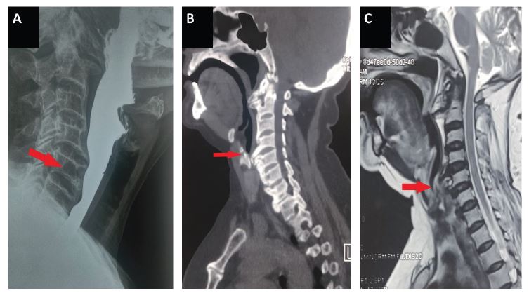
A 61 year old woman with no prior comorbidities presented to our department with symptoms of gradually progressive dysphagia for 2 year duration. Dysphagia was more for solids than liquids. According to the Atkinson Dysphagia severity grading scale, she had Grade 1 dysphagia [3]. She did not have a change in voice, nasal regurgitation, and difficulty in breathing. There was no history of vomiting, heartburnor chest pain. Her vitals were stable, and clinical examination was within normal limits. Barium swallow for the evaluation of dysphagia revealed osteophytes extending from C4-C5 and C5-C6 vertebral junction, causing smooth indentation over the posterior aspect of the esophagus. Further CT scan and MRI of the Neck was done, which showed degenerative changes in the cervical spine with a large anterior osteophyte indenting the esophagus. Upper GI Endoscopy revealed a smooth bulge just above the cricopharynx, with normal esophageal peristalsis. Spine surgeon consultation was obtained, and she was advised conservative management with monitoring. The nature of the disease was explained to the patient and kept in regular follow-up.
Discussion
Dysphagia refers to difficulty in swallowing food, results in abnormal delay or inability to transfer food from the mouth to the stomach. Dysphagia could be oropharyngeal or esophageal, intermittent or progressive, due to mechanical causes or motility disorder [4]. Mechanical dysphagia can be due to intraluminal or external compression of theesophagus. Osteophytes formed at the cervical level due to degenerative changes in old age are a rare cause of dysphagia. Osteophytes are more commonly found at the C5-C6 and C6-C7 levels [5]. The mechanisms of dysphagia secondary to cervical osteophytes previously described in literature were direct compression by osteophytes, pharyngo-esophageal irritation and cricopharyngeal spasm [6]. In an elderly population with dysphagia, a study revealed that approximately 10.6% of the patients had cervical osteophytes as the etiology for dysphagia [7]. Barium swallow study is essential in the diagnosis of obstruction of the esophagus by osteophyte [8]. CT and MRI studies may be needed before planning surgical management. Most of the cases are managed conservatively, but some authors recommend surgical management because of possible progression to acute respiratory distress [9]. Recurrence of symptoms even after surgical resection has been reported previously [10]. The decision to undergo surgery has to be made on individual case to case basis. Our case was managed conservatively after consulting the concerned specialist and kept in regular follow-up to monitor the progression of symptoms.
Conclusion
In elderly patients presenting with mechanical dysphagia, rare causes like an osteophyte compressing the esophagus have to be considered. Management decisions should involve a multidisciplinary team, including a spine surgeon, tailored on a case-by-case basis. All patients on conservative management should be regularly monitored for progression of symptoms.
References
- Howden CW. Management of acid-related disorders in patients with dysphagia. Am J Med. 2004; 117: 44S-48S.
- Lecerf P, Malard O. How to diagnose and treat symptomatic anterior cervical osteophytes? Eur Ann Otorhinolaryngol Head Neck Dis. 2010; 127: 111–6.
- Ogilvie AL, Dronfield MW, Ferguson R, Atkinson M. Palliative intubation of oesophagogastric neoplasms at fibreoptic endoscopy. Gut. 1982; 23: 1060–7.
- Philpott H, Garg M, Tomic D, Balasubramanian S, Sweis R. Dysphagia: Thinking outside the box. World J Gastroenterol. 2017; 23: 6942–51.
- Schmidek HH. Cervical spondylosis. Am Fam Physician. 1986; 33: 89–99.
- Srinivas P, George J. Cervical osteoarthropathy: an unusual cause of dysphagia. Age Ageing. 1999; 28: 321–2.
- Granville LJ, Musson N, Altman R, Silverman M. Anterior cervical osteophytes as a cause of pharyngeal stage dysphagia. J Am Geriatr Soc. 1998; 46: 1003–7.
- Chen YR, Sung K, Tharin S. Symptomatic Anterior Cervical Osteophyte Causing Dysphagia: Case Report, Imaging, and Review of the Literature. Cureus. 2016; 8: e473.
- Maiuri F, Cavallo LM, Corvino S, Teodonno G, Mariniello G. Anterior cervical osteophytes causing dysphagia: Choice of the approach and surgical problems. J Craniovertebr Junction Spine. 2020; 11: 300–9.
- Miyamoto K, Sugiyama S, Hosoe H, Iinuma N, Suzuki Y, Shimizu K. Postsurgical recurrence of osteophytes causing dysphagia in patients with diffuse idiopathic skeletal hyperostosis. Eur Spine J. 2009; 18: 1652–8.