Journal of Gastroenterology Research and Practice
Review Article - Open Access, Volume 5
Portal vein thrombosis: A detailed review of etiology, diagnosis, and treatment
Ali Chand1; Muhammad Ahmad Imran2; Musa Khalil1; Muhammad Hassan1; Qurrat ul ain Iqbal3; Pinky Lohana1; Mudasir Rashid2*
1Department of Medicine, Howard University Hospital, Washington, DC, USA.
2Cancer Center, Howard University Hospital, Washington, DC, USA.
3Department of Medicine, Lahore, Pakistan.
*Corresponding Author : Mudasir Rashid
Cancer Center, Howard University Hospital, Washington, DC, USA.
Email: mudasir.rashid@howard.edu
Received : Jun 25, 2025
Accepted : Jul 23, 2025
Published : Jul 30, 2025
Archived : www.jjgastro.com
Copyright : © Rashid M (2025).
Abstract
Portal Vein Thrombosis (PVT) is a complicated vascular condition resulting from genetic, metabolic, and environmental risk factors. This review underlines current evidence on PVT pathogenesis, clinical management, and emerging therapeutic strategies. Multiple causes of PVT include liver cirrhosis, hepatocellular carcinoma, intra-abdominal infections, and systemic hypercoagulable states, including COVID-19-related thrombosis. Diagnostic challenges persist due to asymptomatic presentations and overlapping imaging findings with other conditions; however, advancements in imaging have improved detection and risk stratification. Anticoagulants remain the cornerstone of treatment, and surgical interventions are reserved for non-responders. Prognosis is influenced by underlying liver disease, thrombus extent, and timely intervention, with cirrhotic PVT linked to higher mortality. Emerging therapies, including miRNA-modified stem cells and radiotherapy for tumor thrombi, show promise but require further validation. Future research must address gaps in genetic pre disposition, optimized DOAC dosing in advanced cirrhosis, and the role of gut microbiota in thrombogenesis. This review highlights the importance of personalized, multidisciplinary approaches to enhance outcomes in this clinically diverse condition.
Keywords: Portal vein thrombosis (PVT); Hypercoagulability; Oral anticoagulants (DOACs); Biomarkers; Gut-liver axis; Thrombolytic therapy.
Citation: Chand A, Imran MA, Khalil M, Hassan M, Rashid M, et al. Portal vein thrombosis: A detailed review of etiology, diagnosis, and treatment. J Gastroenterol Res Pract. 2025; 5(3): 1234.
Introduction
Portal Vein Thrombosis (PVT) is a noteworthy vascular disorder marked by the development of a blood clot in the portal vein and its branches, extending up to the Splenic Vein (SV) and superior mesenteric vein (SMV) [1,2]. It is characterized by partial or complete obstruction of the portal vein, which supplies 3/4th blood to the liver. This obstruction results in compromised hepatic perfusion, potential liver dysfunction and gastrointestinal bleeding [3-5]. This condition is also associated with severe complications such as increased portal venous pressure, variceal bleeding, intestinal ischemia, and liver failure, significantly impacting survival and prognosis [6]. PVT also presents significant challenges in liver transplantation, increasing both perioperative risks and technical difficulties It is most commonly seen in patients with liver cirrhosis, where it often results from portal hypertension, making individuals susceptible to complications like acute esophageal variceal bleeding [7]. In non-cirrhotic cases, PVT often stems from portal and splenic vein thrombosis, leading to variceal bleeding and splenomegaly [8]. PVT is also associated with malignancies, inflammatory conditions, and Myeloproliferative Neoplasms (MPNs), broadening its clinical context [9,10]. Inneonates, PVT is typically asymptomatic and often remains undiagnosed during this early stage of life [11]. Neonatal PVT is recognized as a key factor in the development of portal hypertension [11]. PVT is frequently a symptomatic and often discovered incidentally during routine imaging, particularly during the surveillance of Hepatocellular Carcinoma (HCC) or during hospitalization due to complications related to portal hypertension, such as esophageal variceal bleeding [12]. In cirrhosis, PVT serves as a marker of disease severity and is associated with worsened liver function, portal hypertension, and complications such as esophagogastric varices and ascites [13-15]. PVT is often asymptomatic and typically detected incidentally during routine surveillance imaging of Hepatocellular Carcinoma (HCC) or hospitalization due to complications of portal hypertension [12]. Diagnostic tools such as Doppler ultrasonography are crucial for confirming the diagnosis, as they offer high sensitivity for detecting portal vein occlusion [16]. A holistic overview of Portal Vein Thrombosis (PVT) for clinical research reference and descried in (Figure 1), followed by details sections below:
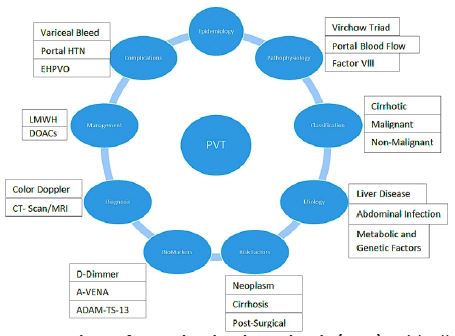
Epidemiological burden
PVT is especially prevalent among patients with advanced liver disease, especially cirrhosis and HCC, with rates ranging from 5% to 26% among those with cirrhosis and up to 40% in HCC cases. The prevalence ranges between 0.6% and 16% among patients with compensated cirrhosis [17-23]. Meta- analyses suggest a pooled prevalence of around 14%, although this figure varies depending on the population and diagnostic techniques used [24-26]. The Neonatal Intensive Care Units (NICU) patients are at higher risk for developing PVT [27]. The incidence of neonatal PVT varies widely, ranging from 1.3% to 43% [27]. Studies also indicate that the incidence of PVT increases in more advanced stages of cirrhosis, with annual rates ranging from 3% to 25% [28]. PVT is considered rare in the population without chronic liver disease, with an incidence of 2-4 per 100,000 individuals [29]. Studies have shown that PVT is responsible for 5% to 10% of all portal hypertension cases in developed countries, while in developing nations, it accounts for upto 33% of cases [8,30,31]. Autopsy studies indicate a prevalence of PVT between 6% to 64%, while studies based on ultrasound report percentages between 5% and 24% [32]. Remove this reference [25,33].
Pathophysiological insights
The development of PVT is a complex, multifactorial process involving Virchow’s Triad [17,34,35]. In cirrhotic patients, the primary contributors are decreased portal blood flow, elevated intrahepatic vascular resistance, along with increased factor VIII levels and diminished protein Clevels [18,36]. The reduction in portal blood flow is particularly pronounced in patients with advanced cirrhosis (Child-Pugh class C) compared to those with milder forms of the disease (Child-Pugh A/B) [37,38]. Genetic factors and systemic conditions, such as endotoxemia, further increase the thrombotic risk [18]. The pathophysiology of cirrhosis involves complex hemostatic alterations due to liver synthetic dysfunction, portal hypertension, and endothelial activation [39]. The mechanism of PVT in cirrhosis is distinct from other thrombotic conditions, such as Deep Vein Thrombosis (DVT) or Pulmonary Embolism (PE) [16,40]. Unlike the thrombi found in DVT or PE, which primarily consist of fibrin, platelets, and red blood cells, portal vein thrombi are mainly composed of intimal hyperplasia [41,42]. This suggests that terms like “portal vein obstruction” or “portal vein stenosis” may be more appropriate in describing PVT in cirrhosis [16,43]. Further contributing factors to PVT include portal hypertension, endothelial injury, the presence of esophageal varices, and a history of variceal endoscopic treatments [32]. The prothrombotic nature of PVT is also linked to the involvement of Neutrophil Extracellular Traps (NETs), which promote clot formation. NETs, composed of DNA and histones, provide as caffold for thrombus development and possess strong procoagulant activity [45,46]. Additionally, propranolol, a Nonselective β-Blocker (NSBB) commonly used in cirrhosis management, has been shown to enhance NET formation by increasing the production of Reactive Oxygen Species (ROS) and NADPH oxidase activity [47,48]. This highlights the paradoxical role of NSBBs in potentially contributing to PVT despite their effectiveness in reducing variceal bleeding [38].
Classification of PVT
“PVT can be classified into cirrhotic PVT, malignant thrombosis, and non-malignant-non-cirrhotic PVT.” [35]. This line provides a basic classification framework for PVT, which is essential in understanding its different types based on etiology (detail given in Table 1).
Table 1: Classification of PVT.
| Classification | Description | Ref |
|---|---|---|
| Cirrhotic PVT | Liver cirrhosis is responsible for about 33%of all PVT cases. The elevated incidence of PVT in advanced cirrhosis is attributed to theVirchow’s triad* Among patients with compensated cirrhosis, the prevalence isaround 1%. | [49,50] |
| Malignant Thrombosis: | Malignant PVT is typically linked to the advancement of underlying malignancies. According to somestudies, PVT wasidentified in 14.3% ofpatients with primary liver cancer accompanied by cirrhosis, and in 11.5% ofthose diagnosed with pancreatic cancer. | [35,51,52] |
| Non-malignant-non-cirrhoticPVT: | Non-malignant and non-cirrhotic PVTis attributed to prothrombogenetic conditions. For example, 20-50% of thepatients with NMNC PVT have reported myeloproliferative neoplasias. Similarly, In a meta-analysis of 855 patients with PVT, 30% of the patientshad underlying myeloproliferative neoplasms. | [53,54] |
Portal Vein Thrombosis (PVT). Virchow's triad* (hypercoagulability, endothelial injury, stasis)
Etiological factors and risk contributors
PVT can arise from various etiological factors, encompassing both inherited and acquired conditions. These factors disrupt normal blood flow and lead to thrombogenesis in the portal venous system. The primary causes of PVT include liver diseases (such as cirrhosis and non- alcoholic steatohepatitis), infections, abdominal surgeries, and systemic inflammatory states. Other etiological factors include:
Schistosomiasis-induced PVT: The pathophysiology of PVT in schistosomiasis is associated with chronic pelvic adhesions and inflammation, which contribute to thrombogenesis [55]. Risk factors for schistosomiasis-induced PVT include cirrhosis, systemic inflammation, reduced Portal Vein Velocity (PVV), wider Portal Vein Diameter (PVD), and the presence of Gastroesophageal Varices (GOV) [16,56,57].
Intra-abdominal infections: Intra-abdominal infections are significant contributors to PVT, with pylephlebitis (suppurative thrombophlebitis of the portal vein) being a key complication. Conditions such as appendicitis, diverticulitis, Inflammatory Bowel Disease (IBD), cholecystitis, and pancreatitis have been implicated in the development of PVT. Among these, diverticulitis is a notable risk factor, with an incidence of approximately 3% in colonic diverticulitis [58-67].
COVID-19 and thrombosis: The hypercoagulable state induced by COVID-19 significantly increases the risk of thrombosis, including PVT and mesenteric vein thrombosis. COVID-19 induces a cytokine storm and endothelial dysfunction, both of which promote thrombosis in unusual sites [68]. Vaccine-induced Immune Thrombotic Thrombocytopenia (VITT), which has been associated with the COVID-19 vaccines, can sometimes result in thrombosis within weeks of vaccination [69].
Systemic infections and thrombosis: Systemic Infection associated inflammatory responses can lead to a hypercoagulable state, thereby increasing the risk of Venous Thromboembolism (VTE), including PVT. Infections such as acute toxoplasmosis and bacterial infections with hypervirulent strains of Klebsiella pneumoniae contribute to thrombosis formation through inflammatory mediators and Disseminated Intravascular Coagulation (DIC) [70-75].
Inflammatory bowel disease (IBD): Patients with IBD, particularly those with Ulcerative Colitis (UC) and Crohn’s Disease (CD), have a significantly higher risk of developing PVT and other VTE-related conditions. A nationwide study has shown a 20% increase in VTE-related hospitalizations among IBD patients, with UC linked to an elevated risk of thrombosis [76,77]. This underscores the need for preventive strategies in IBD patients to reduce thrombosis risk.
Cirrhosis and liver dysfunction: Cirrhotic patients are in a hyper coagulable state due to elevated von Wille brand factor (vWF) and reduced ADAMTS-13, leading to platelet aggregation and thrombus formation [78]. Factor VIII and deficiencies in Protein C and S also contribute to increased thrombin generation and PVT risk [79-81]. The presence of cirrhosis complicates the diagnosis of PVT, with debates surrounding its role in exacerbating cirrhosis progression or merely reflecting disease severity [82].
Liver hypoplasia: Liver hypoplasia, a rare condition leading to elevated intrahepatic portal venous pressure, can contribute to PVT. This is particularly relevant for diagnosis in patients unundergoing liver transplantation or other surgical procedures [83-85].
Clomiphene citrate use: Clomiphene Citrate (CC), a drug commonly used for ovulation induction and male infertility treatment, has been associated with an increased risk of thrombosis, including PVT and Splanchnic Vein Thrombosis (SMVT). The drug’s effect on hormonal levels and coagulation pathways may increase the likely hood of venous thrombo embolism. Clinical case studies have demonstrated that patients using clomiphene, with no other apparent risk factors, developed PVT and SMVT, suggesting a potential causative role of the medication [86-88].
Metabolic comorbidities: Patients with Non-Alcoholic Fatty Liver Disease (NAFLD) area tan elevated risk for developing PVT due to associated metabolic comorbidities such as obesity, type 2 diabetes, and dyslipidemia [89]. Additionally, individuals with COVID-19, particularly those with severe disease or elevated D-dimer and Lactated Hydrogenase (LDH) levels, have been found to have an increased incidence of thromboembolic events, including PVT [90,91].
Genetic factors: Genetic predis positions significantly influence the risk of PVT. Mutations such as Factor V Leiden and prothrombin G20210A have been implicated in increasing thrombotic risk in affected individuals [92,93]. Additionally, the MTHFR TT genotype is associated with an earlier onset of PVT due to elevated Homocysteine (HC) levels, which contribute to oxidative damage and thrombosis [94,95].
Hypercoagulable states: Hypercoagulability during pregnancy significantly increases the risk of PVT, especially when compounded by obesity (BMI >30 kg/m²). Pregnancy-induced changes, such as increased coagulation activity after 28 weeks of gestation, contribute to a heightened thromboembolic risk [96-99].
Risk factors for portal vein thrombosis
Myelo proliferative neoplasms and thrombophilia: Clonal hematopoiesis in myeloproliferative neoplasms (MPNs) like polycythemia vera and essential thrombocythemia significantly raises thrombotic risks, including splanchnic vein thrombosis. The JAK2V617F mutation is particularly linked to PVT [9,100,101]. Thrombophilic disorders and low ADAMTS13 activity further exacerbate thrombotic tendencies [16,102].
Post-surgical and environmental risks: PVT has been documented following bariatric surgeries such as sleeve gastrectomy, where the incidence reaches 0.5%, attributable to hypercoagulability and inflammation [103]. Splenectomy, often performed to manage portal hypertension, is another notable risk factor, with PVT incidence rates post-surgery ranging between 18.9% and 57% [104,105].
Metabolic and virological Factors: Metabolic conditions, including advanced cirrhosis and virological diseases like hepatitis B, heighten PVT risk. Hepatic tuberculosis, though rare, can directly affect the portal vein, leading to thrombosis [106,107]. Sustained virological response via direct-acting antivirals reduces hypercoagulability in cirrhotic patients but does not entirely eliminate the risk in advanced cases [108].
Risk factors in neonates: The placement of Umbilical Venous Catheters (UVCs) in neonates has been identified as a significant risk factor for PVT. This triggers bacterial dissemination, which activates the coagulation cascade and leads to thrombus formation [109-110]. Malpositioned UVCs contribute to vessel wall irritation and clot formation. Studies have demonstrated that a notable proportion of neonates with UVCs develop PVT, and prolonged follow-up is essential for children with this risk factor [111,112].
Cancer-related risk factors: Hepatocellular Carcinoma (HCC) and cirrhosis contribute significantly to the development of PVT due to a combination of tumor-induced hypercoagulability, liver inflammation, and altered coagulation dynamics. Tumor-induced cytokine release, platelet activation, and extracellular matrix remodeling further promote thrombosis [113-116]. Additionally, cancer stem cells, particularly those expressing EpCAM and CD133, are linked to PVT and metastasis [117,118].
Other risk factors: Alterations in gut microbiota, such as reduced Bacteroides abundance, have been linked to PVT progression in cirrhotic patients, with interventions aimed at restoring the microbiota showing promise in improving outcomes [119]. Additional risk factors for PVT include older age, splenomegaly, ascites, elevated INR, low albumin levels, and high MELD scores [120-122].
Biomarkers and molecular predictors
The identification of reliable biomarkers for predicting Portal Vein Thrombosis (PVT) is essential for early detection, risk stratification, and monitoring disease progression. Several clinical and laboratory biomarkers have shown promise in aiding the diagnosis and prognosis of PVT in various patient populations (detail given in Table 2).
Table 2: Biomarkers in portal vein thrombosis.
| Biomarkers | Description | Ref |
|---|---|---|
| D-Dimer: | Higher levelsof D- dimer and a larger portalvein diameter havebeen associated with an increased risk of thrombus formation, whereas lower platelet counts may reflect acompromised hemostatic response | [123,124] |
| A-VENA Criteria and PIVKA-II: | The A-VENA criteria, which incorporate clinical variables andbiomarker data, have proven useful inidentifying high-risk individuals. One key biomarker, Protein Induced by Vitamin K Absence or Antagonist II (PIVKA-II), has been shownto help in identifyingpatients at high risk of developing PVT, particularly in those with liverdisease | [125,126] |
| ADAMTS-13and VWF | Lowlevels of ADAMTS-13 activity (<18.8%) and an imbalanced ADAMTS-13/VWF# ratio have been identified as strong predictors of PVT, especially in cirrhotic patients. A ratio of ADAMTS-13 to VWF lessthan 0.4 has been suggested as a reliable marker for PVT prediction. | [102,127,128] |
| Systemic Inflammatory Markers | Inflammatory indices, such as the Albumin-to-Neutrophil RatioIndex (ANRI), Platelet-Lymphocyte Ratio(PLR), and Neutrophil- Lymphocyte Ratio (NLR), have emerged as useful markers inpredicting PVT. Elevated levels of these markers are associated with an increased risk of PVT,particularly incirrhotic and critically ill patients | [124,129,130] |
| IntestinalMicrobiota | Emerging research has highlighted the role of the gut-liver axis and intestinal microbiota in the development of thrombosis, including PVT. Imbalances in the gutflora may contribute to thrombogenesis, influencing coagulation pathways and exacerbating the hypercoagulable state. | [131] |
| Validation of Biomarkers | While these biomarkers are promising, largerprospective studies are needed to validate theirclinical utility. Standardization and reproducibility of these markers across different patient populations and clinical settings are crucial for their widespread adoption in clinical practice | [124] |
Table 3: Diagnostic methods in portal vein thrombosis.
| Methods | Description | Ref |
|---|---|---|
| Color Doppler Ultrasound: | Color Doppler ultrasound is the first-line diagnostic tool for PVT due to itsaccessibility and non-invasive nature. Thistechnique is particularlyeffective for detecting the presence of thrombus and assessing portal veinpatency. It typically shows high-levelechoes, non-visualization of the portalvein, and serpiginous vascular channels in cases of thrombosis. Sensitivity ranges from 60% to100%, depending on the operator’s expertise, and it is especially useful fordetecting complications like portal hypertension,varices, and splenomegaly. However, Doppler ultrasonography may miss early-stage PVT, especially in cirrhotic patients. | [120,132-134] |
| Contrast- enhanced CT and MRI scans: | Contrast-enhancedCT and MRI scans are preferred for confirming PVT, as they provide superiorsensitivity and specificity compared to ultrasound. Theyare also essential for detecting complications of thrombosis, whichcan occur within6–20 days. MRI, in particular, has demonstratedhigh sensitivity (100%) and specificity (98%) in detecting PVT.(PMID:1853809, PMID: 8816536) | [135-137] |
| Contrast- enhanced ultrasound (CEUS) | CEUShas become an increasingly valuable tool for diagnosing PVT, particularly indifferentiating between benign and malignantthrombi. CEUS is particularly usefulfor detecting thrombi in patients with hepatocellular carcinoma (HCC), depicting rapidarterial- phase hyper-enhancement and slow portalvenous washout. Studieshave shown CEUStohave100%sensitivityand98%specificityfor tumor-relatedPVT.However, the diagnosticaccuracy of CEUS can vary based on operator expertise. | [138,139] |
| EGD | Esophagogastroduodenoscopy (EGD)plays a critical role in evaluating complications of PVT,particularly for variceal detection. Theprocedure allows for the direct visualization of gastro esophageal varices,which are commonly associated with portal hypertensionresulting from PVT. | [140] |
| Contrast- enhancedCT-Scan forPylephlebitis | Pylephlebitis, an infection of the portalvein, presents withnonspecific symptoms, makingits diagnosis challenging. Contrast- enhancedCT is the imaging modality of choice for detecting pylephlebitis, revealingportal vein dilatation, thrombus, wall thickening,and complications such as hepatic abscess or mesenteric ischemia. | [141,142] |
| Portal-phase imaging | Portal-phaseimaging, particularly with CT and MRI, is crucial for identifying thrombosisand its complications, such as hepatic abscesses or intestinal ischemia. However, definitive evidence of infectious thrombic an be challenging to obtain, underscoring the diagnosticcomplexities involved in PVT management. | [62,72,143] |
| Imaging for Hepatic TB | HepaticTuberculosis (TB) can present with imaging findings that overlap with PVT.Ultrasound and CT may suggest hepatic TB, but theirlack of specificity can lead to diagnostic challenges. Miliary lesions on CT appearas micro abscesses, while local hepatic TB manifests as larger nodules that may resemble neoplastic orgranulomatous diseases. Histopathological or bacteriological confirmation is often required for adefinitive diagnosis of hepatic TB, with liver biopsy revealing granulomas. | [144,145] |
| PCRTesting for Hepatic TB | PCR testingtargeting the IS6110sequence has demonstrated higher sensitivity (86%)and specificity (96%)for diagnosing hepaticTB. | [146] |
| Portal-phase imaging | Portal-phase imaging, particularly with CT and MRI,is crucial for identifying thrombosis and its complications, such as hepaticabscesses or intestinal ischemia. | [62.65] |
Table 4: Management of PVT.
| Management type | Description | Ref |
|---|---|---|
| Thrombolysis andSurgical Intervention | ||
| Thrombolysis | Thrombolytictherapy (e.g., urokinase, streptokinase, Tissue Plasminogen Activator [TPA])can be considered in acute cases within14–30 days of diagnosis. Mechanical thrombectomy, often used during bowelinfarction surgeries, is still not definitivelyeffective, as recurrent thrombosis and the need for further interventions arecommon. | [159,160] |
| Surgical Intervention | Surgical thrombectomy is reserved forcases involving bowelinfarction but carries a high riskof re-thrombosis. Forsevere or refractory cases,consider transjugular intrahepatic portosystemic shunt (TIPS) or livertransplantation. | [161-163] |
| Endoscopic varicealligation (EVL) | Endoscopic Variceal Ligation (EVL) and beta blockersareess entail for primary and secondary prophylaxis of acute variceal bleeding in Extrahepatic Portal Vein Obstruction (EHPVO) | [164] |
| Anticoagulation Post-Splenectomy | Anticoagulation is crucial after splenectomy to prevent PVT complications, particularly in patients withesophagealvarices. | [103] |
| Antibiotic Therapy | ||
| Broad Spectrum IVAntibiotics | Initiate broad-spectrum intravenous antibiotics immediately upon diagnosis and transition to oral antibiotics after two weeksfor a totaltreatment duration of six weeks.Early antibiotic intervention is crucial for reducing complications and preventing progression | [65,148,165] |
| Management of Specific Conditions | ||
| Splenic Vein Thrombosis (SVT) | In cases of SVT secondary to acute pancreatitis, anticoagulation is usedto aid recanalization, though it doesnot significantly affectmortality. | [166] |
| MyeloproliferativeNeoplasms (MPNs): | Long-term anti coagulation with DOACs is effective, and adding aspirin may help preventrecurrence | [9,167,168] |
| Hepatic Tuberculosis(TB): | Combine Anti-Tuberculosis Therapy (ATT) withanticoagulation, with DOACspreferred due to their safetyand ease of use. | [104,168] |
| COVID 19 Considerations | ||
| LMWH Prophylaxis: | Prophylactic LMWH reduces thromboembolic events in COVID-19 patients, though its impacton PVT incidence is unclear. | [90,169]. |
| Anticoagulation in Hemophilia | ||
| Extended Half-Life FIXConcentrates: | Used in hemophilic patients with PVT to maintain coagulation balance and reducebleeding risks. | [170-172] |
| Mesenchymal Stem Cells (MSCs): | miRNA-25-3p-modified humanumbilical cord MSCspromote endothelial repairby enhancing cellproliferation, migration, and angiogenesis, potentially aiding inPVT management. | [173-175] |
| Exosomal miRNAs: | MSC-derived exosomes have anti-inflammatory and tissue repairproperties, reducing thrombotic events and promoting endothelial function via exosomal miRNAs like miRNA-126-3p andmiRNA-342-3p. | [176-181] |
| Therapeutic Potential: | The PTEN/KLF4/AKT/ERK1/2 signalingpathway mediatesthebeneficialeffectsofmiRNA-25-3p-modified hucMSCs, underscoringtheir potential as a therapy for PVT. | [182,183] |
| Anti-coagulationin Pylephlebitis | ||
| Heparin andDOACs: | Heparin and DOACs (e.g.,apixaban) are effective for managing pylephlebitis, with DOACs preferred due to theirlower bleeding risk. Optimalduration remains unclear | [63] |
| Antibiotic Selection: | Antibiotics shouldaccount for the infection’s originand bacterial resistance, with ceftriaxone and ampicillin-sulbactam being successful, while cefmetazoleshould be avoided | [184] |
| Emerging Therapies | ||
| Systemic Therapies in HCC | Combination therapies like atezolizumab andbevacizumab provide superior survival outcomes compared to sorafenib in advancedHCC with PVT. Studies also suggest the effectiveness of Immune CheckpointInhibitors (ICIs). | [185-187] |
| Trans-arterial radioembolization (TARE) | Emerging therapies, suchas TARE with Yttrium-90 (Y-90)and Stereotactic BodyRadiation Therapy (SBRT), offer better tumor control while preserving livertissue, showing potential in improving outcomes in PVTT management. | [188-190] |
| Portal Vein Recanalization (PVR) in Children | PVRis an emerging alternative to traditional surgical methods likeMeso-Rex by pass in children withEHPVT, offering the advantage of restoring the native portalvenous system and avoiding complications associated with portosystemic shunts. Predictorsof success for PVR remain unclear, butit avoids complications like hepaticence phalopathy | [191,192] |
Diagnostic approaches
Accurate and timely diagnosis of PVT is crucial for appropriate clinical management. Several diagnostic techniques are employed to detect PVT and assess its severity, with imaging modalities playing a central role in confirming the diagnosis. These techniques include ultrasound, contrast-enhanced imaging (CT and MRI), and advanced diagnostic methods that help identify thrombus presence, associated complications, and underlying conditions such as cirrhosis or malignancy (detail given in Table 3) and diagnostic challenges in Portal Vein Thrombosis have been described in Supplementary Table 1.
Initial management strategies
The management of PVT involves a comprehensive approach tailored to the underlying cause, severity, and patient-specific factors. Initial treatment typically includes rapid anticoagulation with Low Molecular Weight Heparin (LMWH) or unfractionated heparin, followed by a prolonged course (≥6 months) to prevent thrombus progression. Once stabilized, patient is transitioned to oral anticoagulants like war far in (targetINR:2-3) or Directoral Anti Coagulants (DOACs) such as apixaban, rivaroxaban, or dabigatran [65,147-151]. DOACs often preferred due to their safety profile and ease of use. In cases where anticoagulation fails or is contraindicated, interventions like Trans-jugular Intrahepatic Portosystemic Shunt (TIPS) or surgical thrombectomy may be necessary. Adjunct therapies such as endoscopic variceal ligation and beta blockers are essential for preventing variceal bleeding [152-158] (Table 4).
Prognosis and outcome predictors
Impact of anticoagulation and underlying conditions: The prognosis of PVT is majorly influenced by the timely initiation of anti-coagulation therapy and management of underlying conditions. In cirrhotic patients without anticoagulation therapy, approximately 22% experience progression of PVT, while 77.7% have stable or improved conditions. Notably, 29.3% of cases experience regression of PVT, but complete recanalization occurs in only 10.4%, and recurrence is observed in 24% of patients [193].
Effect of liver disease and coexisting conditions: The underlying liver disease is a key factor in determining prognosis. Cirrhotic patients with PVT experience a significant decrease in two-year survival rates, primarily due to impaired liver function and associated complications [194]. In contrast, patients with Non-Alcoholic Fatty Liver Disease (NAFLD) related PVT generally have a better prognosis when treated with anticoagulation therapy. The NAFLD patients showed complete recanalization within six months without long-term complications with the use of edoxaban to prevent recurrence [195]. Factors such as a higher MELD score and the presence of ascites are linked to worse outcomes in cirrhotic patients with PVT [193]. For non-cirrhotic, non- malignant PVT, the 1-year mortality rate is lower (8%) compared to malignancy- or cirrhosis- associated PVT, which has a higher 1-year mortality rate (~26%) [196].
Acute PVT with intestinal ischemia: Acute PVT complicated by intestinal ischemia has a particularly poor prognosis, with mortality rates ranging from 20% to 50% [30,197]. Early intervention and anticoagulation therapy can improve survival rates in these cases, highlighting the importance of prompt diagnosis and management.
PVT and venous thromboembolism (VTE): PVT is strongly associated with Venous Thromboembolism (VTE), Including Pulmonary Embolism (PE), with an increased risk of VTE in PVT patients, particularly in those with idiopathic PVT [198]. The risk of PE is concerning due to its high mortality rates, especially when associated with cardiac arrest, as 70% of fatal cardiac arrests occur within the first hour of PE onset [199,200]. Therefore, early preventive measures and monitoring for VTE and PE in PVT patients are critical to improving prognosis.
Prophylaxis and long-term monitoring: For cirrhotic patients, prophylactic anticoagulation therapy may decrease the incidence of PVT and improve long-term outcomes [201]. Long-term anticoagulation therapy has been shown to reduce the frequency of variceal bleeding episodes, improve outcomes by lowering microvascular thrombosis, and reduce portal vein pressure [202]. However, patients with cirrhosis and low platelet counts (below 50,000/mL) may be at higher risk for bleeding, with major bleeding rates reaching up to 9% [150,203].
Extrahepatic portal vein obstruction (EHPVO): In patients with Extrahepatic Portal Vein Obstruction (EHPVO), long-term survival can be improved with individualized care, including primary and secondary prophylaxis for variceal bleeding [140]. Proximal splenorenal shunts offer an 80% survival rate, while Rex surgery boasts nearly 100% long-term survival [140]. Post-surgical monitoring is critical to detect complications such as variceal recurrence and portosystemic encephalopathy.
Vascular invasionin hepatocellular carcinoma (HCC): Vascular invasion, particularly in HCC with PVT, significantly impacts survival. Patients with vascular invasion (Vp1-Vp4) exhibit progressively worse survival rates, with Child-Pugh A patients undergoing hepatic resection showing median survival rates of 34 months [204]. Survival decreases as the degree of vascular invasion increases (Vp1: 42.7%, Vp2: 25.2%, Vp3: 22.3%, Vp4: 9.8%) [204].
Pylephlebitis and long-term monitoring : Pylephlebitis, when left untreated, can lead to severe complications such as hepatic abscess, mesenteric ischemia, portal hypertension, and pulmonary embolism, which significantly worsen prognosis. Regular follow-up imaging is essential to monitor thrombus resolution and prevent complications [63,205]. Aggressive management, including anticoagulation and long-termmonitoring, is critical for improving patient out comes and reducing morbidity and mortality associated with this condition [148].
Portal hypertension (PHT): Portal hypertension plays a crucial role in the prognosis of PVTT, exacerbating complications such as refractory ascites and esophagogastric varices, which can lead to gastrointestinal bleeding. Managing PHT through interventions like portal vein stenting and radio Frequency Ablation (RFA) can improve survival outcomes in PVTT patients by reducing the risk of bleeding and worsening liver dysfunction [206].
Survival and prognostic indicators in PVT: Survival in PVT is significantly influenced by the extent of thrombosis and the presence of vascular invasion, with survival times being particularly short when the main portal vein is involved (MPVTT). Median survival in such cases ranges from 2.7 to 4 months [185,207,208]. These high lights the importance of early detection, aggressive management, and personalized treatment strategies to improve prognosis, especially in high-risk patients.
Complications and clinical challenges
Variceal re-hemorrhage: Chronic PVT significantly heightens the risk of variceal re-hemorrhage [209]. Patients with cirrhosis and cavernous transformation of the portal vein generally have better outcomes, including lower mortality rates, compared to those without cavernous transformation [210]. A recent study revealed that TIPS is more effective than anticoagulation alone in achieving portal vein recanalization and improving survival, with reduced rates of variceal rebleeding and refractory ascites in the TIPS group [211].
Portal hypertension and its effects: Portal hypertension in PVT can lead to the development of varices, splenomegaly, and hypersplenism, all of which require careful management [212,213].
Extrahepatic portal vein obstruction (EHPVO): In patients with Extrahepatic Portal Vein Obstruction (EHPVO), variceal bleeding, particularly from ectopicvarices, is a significant clinical challenge [214,215].
Septicemia and bacterial translocation: A rare but serious complication in PVT patients is Clostridium paraputrificum septicemia, typically observed in immunocompromised individuals or those with gastrointestinal pathology [216,217]. In cases of ischemic bowel disease, the disruption of the mucosal barrier facilitates bacterial translocation into the bloodstream [218]. The introduction of MALDI-TOF mass spectrometry has enhanced the detection of anaerobic infections, improving diagnostic accuracy in these cases [219,220].
Complications in neonates: Inneonates, long-term complications of PVT include liver lobe a trophy, splenomegaly, and portal hypertension, necessitating ongoing monitoring [27].
Future direction and research gap
Further investigation is needed to explore the role of coagulation markers and portal vein velocity in identifying cirrhotic patients at risk for PVT, especially in those who achieve Sustained Virological Response (SVR) [42,221,222]. Focused research into diagnostic advancements, targeted therapies, and optimized anticoagulation protocols is essential to improve outcomes in PVT management [42]. Additionally, the potential role of NSBBs in promoting PVT via NETs and neutrophil activity in cirrhosis warrants further exploration [42,221,222]. Similarly, future studies should also investigate the mechanisms behind clomiphene-associated thrombosis and enhance management strategies for cirrhotic PVT and drug-related thromboembolic events. The limitations of current studies include small sample sizes, which affect the generalizability of findings. The absence of comparison across different disease stages restricts insights into how disease progression influences treatment outcomes. There prospective nature of many studies and single-center settings introduces potential biases. To address these issues, future prospective clinical trials with larger, multicenter populations are needed to validate findings, biomarkers, optimize thrombolytic regimens, and explore the role of gut-liver interactions in PVT development along with the efficacy of combined therapies for PVTT in HCC [102,131,159,206].
Research into miRNA-modified human umbilical cord-derived mesenchymal stem cells (hucMSCs) holds promise for endothelial repair and vascular health [173,174]. Future studies should explore the molecular mechanisms behind miRNA-modified therapies and their broader applications in vascular diseases [180,181]. Similarly, genetic predis position, especially MTHFR genotypes, highlights the need for early screening and personalized therapies for PVT [94,95]. While radiotherapy, including Stereotactic Radiotherapy (SRT) and hypo-fractionated radiotherapy, shows potential for managing PVTT in HCC, challenges remain indetermining the optimal radiation dose and fractionation schedule for External Beam Radiotherapy (EBRT) and SRT. Further studies are necessary to establish the best anticoagulation regimens and minimize side effects to improve therapeutic outcomes for PVT.
Conclusion
PVT is a complex thrombotic condition influenced by a range of genetic, metabolic, and environmental risk factors. Key contributors include hormonal therapies like clomiphene citrate, underlying comorbidities such as obesity and NAFLD, genetic predispositions, and surgical procedures. Additionally, cancer, particularly HCC, liver cirrhosis, infections, and mechanical factors such as bariatric surgery, play significant roles in the pathogenesis of PVT. Advances in the understanding of infections, gut microbiota, micro-vesicles, and cancer stem cells offer opportunities to improve diagnostic tools and therapeutic strategies, thereby reducing thrombotic risk and enhancing patient outcomes [154,223,224]. The treatment of Non-Cirrhotic PVT (NCPVT) primarily includes anticoagulation therapy, with Direct Oral Anticoagulants (DOACs) becoming the preferred choice due to their safety and convenience [154]. Endovascular interventions are increasingly utilized in refractory cases, while careful management of portal hypertensive complications remains essential, especially in chronic PVT [223,224]. The complex and multifactorial nature of PVT demands a thorough and individualized approach to diagnosis and treatment. Ongoing research is needed to better understand the molecular basis of PVT, refine management strategies, and develop predictive models to improve patient outcomes. The development of more effective therapies, coupled with early detection and intervention, will be key to addressing the challenges posed by PVT in both cirrhotic and non-cirrhotic patients [59].
References
- De Stefano V, Martinelli I. Splanchnic vein thrombosis: clinical presentation, risk factors and treatment. Intern Emerg Med. 2010; 5: 487–494.
- Valla DC, Condat B. Portal vein thrombosis in adults: pathophysiology, pathogenesis and management. J Hepatol. 2000; 32: 865–871.
- Martin A, Struble S, Prado A, Robinson J, Goddard J, Smith T. A case of portal vein thrombosis in a patient with methylenetetrahydrofolate reductase A1298C polymorphism. Cureus. 2022; 14: e21743.
- Manzano-Robleda Mdel C, Barranco-Fragoso B, Uribe M, Mendez-Sanchez N. Portal vein thrombosis: what is new? Ann Hepatol. 2015; 14: 20–27.
- Gioia S, Nardelli S, Ridola L, Riggio O. Causes and management of non-cirrhotic portal hypertension. Curr Gastroenterol Rep. 2020; 22: 56.
- Stine JG, Shah PM, Cornella SL, Rudnick SR, Ghabril MS, Stukenborg GJ, Northup PG. Portal vein thrombosis, mortality and hepatic decompensation in patients with cirrhosis: a meta-analysis. World J Hepatol. 2015; 7: 2774–2780.
- In: Acute upper gastrointestinal bleeding in over 16s: management. London; 2016.
- Valla DC, Condat B, Lebrec D. Spectrum of portal vein thrombosis in the West. J Gastroenterol Hepatol. 2002; 17 Suppl 3: S224–227.
- Tremblay D, Winters A, Beckman JD, Naymagon L, Patel R, Mascarenhas J, Schiano TD. Splanchnic vein thrombosis associated with myeloproliferative neoplasms. Thromb Res. 2022; 218: 8–16.
- Alvarez-Larran A, Pereira A, Magaz M, et al. Natural history of polycythemia vera and essential thrombocythemia presenting with splanchnic vein thrombosis. Ann Hematol. 2020; 99: 791–798.
- Williams S, Chan AK. Neonatal portal vein thrombosis: diagnosis and management. Semin Fetal Neonatal Med. 2011; 16: 329–339.
- Allaire M, Thabut D. Reply: Portal hypertension and variceal bleeding in patients with liver cancer: evidence gaps for prevention and management. Hepatology. 2024; 79: E3.
- Yoshida H, Shimizu T, Yoshioka M, Taniai N. Management of portal hypertension based on portal hemodynamics. Hepatol Res. 2021; 51: 251–262.
- Tadokoro T, Morishita A, Masaki T. Diagnosis and therapeutic management of liver fibrosis by microRNA. Int J Mol Sci. 2021; 22: Article 15.
- Amitrano L, Guardascione MA, Brancaccio V, et al. Risk factors and clinical presentation of portal vein thrombosis in patients with liver cirrhosis. J Hepatol. 2004; 40: 736–741.
- Turon F, Driever EG, Baiges A, et al. Predicting portal thrombosis in cirrhosis: a prospective study of clinical, ultrasonographic and hemostatic factors. J Hepatol. 2021; 75: 1367–1376.
- Costache RS, Dragomirica AS, Dumitras EA, et al. Portal vein thrombosis: a concise review (Review). Exp Ther Med. 2021; 22: 759.
- Mantaka A, Augoustaki A, Kouroumalis EA, Samonakis DN. Portal vein thrombosis in cirrhosis: diagnosis, natural history, and therapeutic challenges. Ann Gastroenterol. 2018; 31: 315–329.
- Luo X, Wang W, Fan X, et al. Short-term outcome of patients with cirrhosis and concurrent portal cavernoma presenting with acute variceal bleeding. Can J Gastroenterol Hepatol. 2018; 2018: 9491856.
- Urhut MC, Sandulescu LD, Ciocalteu A, Cazacu SM, Danoiu S. The clinical value of multimodal ultrasound for the differential diagnosis of hepatocellular carcinoma from other liver tumors in relation to histopathology. Diagnostics (Basel). 2023; 13: Article 20.
- Intagliata NM, Argo CK, Stine JG, et al. Concepts and controversies in haemostasis and thrombosis associated with liver disease: proceedings of the 7th International Coagulation in Liver Disease Conference. Thromb Haemost. 2018; 118: 1491–1506.
- Ponziani FR, Zocco MA, Garcovich M, et al. What we should know about portal vein thrombosis in cirrhotic patients: a changing perspective. World J Gastroenterol. 2012; 18: 5014–5020.
- Cerrito L, Annicchiarico BE, Iezzi R, et al. Treatment of hepatocellular carcinoma in patients with portal vein tumor thrombosis: beyond the known frontiers. World J Gastroenterol. 2019; 25: 4360–4382.
- Pan J, Wang L, Gao F, et al. Epidemiology of portal vein thrombosis in liver cirrhosis: a systematic review and meta-analysis. Eur J Intern Med. 2022; 104: 21–32.
- Tsochatzis EA, Senzolo M, Germani G, et al. Systematic review: portal vein thrombosis in cirrhosis. Aliment Pharmacol Ther. 2010; 31: 366–374.
- Nery F, Chevret S, Condat B, et al. Causes and consequences of portal vein thrombosis in 1,243 patients with cirrhosis: results of a longitudinal study. Hepatology. 2015; 61: 660–667.
- Morag I, Epelman M, Daneman A, et al. Portal vein thrombosis in the neonate: risk factors, course, and outcome. J Pediatr. 2006; 148: 735–739.
- Senzolo M, Garcia-Tsao G, Garcia-Pagan JC. Current knowledge and management of portal vein thrombosis in cirrhosis. J Hepatol. 2021; 75: 442–453.
- Ageno W, Dentali F, Pomero F, et al. Incidence rates and case fatality rates of portal vein thrombosis and Budd-Chiari syndrome. Thromb Haemost. 2017; 117: 794–800.
- Ponziani FR, Zocco MA, Campanale C, et al. Portal vein thrombosis: insight into physiopathology, diagnosis, and treatment. World J Gastroenterol. 2010; 16: 143–155.
- Prakash S, Bies J, Hassan M, et al. Portal vein thrombosis in cirrhosis: a literature review. Front Med (Lausanne). 2023; 10: 1134801.
- Fimognari FL, Violi F. Portal vein thrombosis in liver cirrhosis. Intern Emerg Med. 2008; 3: 213–218.
- Francoz C, Valla D, Durand F. Portal vein thrombosis, cirrhosis, and liver transplantation. J Hepatol. 2012; 57: 203–212.
- Hoekstra J, Janssen HL. Vascular liver disorders (II): portal vein thrombosis. Neth J Med. 2009; 67: 46–53.
- Trebicka J, Strassburg CP. Etiology and complications of portal vein thrombosis. Viszeralmedizin. 2014; 30: 375–380.
- Kumar A, Sharma P, Arora A. Review article: portal vein obstruction—epidemiology, pathogenesis, natural history, prognosis and treatment. Aliment Pharmacol Ther. 2015; 41: 276–292.
- Arroyo AB, Fernandez-Perez MP, Del Monte A, et al. miR-146a is a pivotal regulator of neutrophil extracellular trap formation promoting thrombosis. Haematologica. 2021; 106: 1636–1646.
- Noubouossie DF, Reeves BN, Strahl BD, Key NS. Neutrophils: back in the thrombosis spotlight. Blood. 2019; 133: 2186–2197.
- Lisman T, Hernandez-Gea V, Magnusson M, et al. The concept of rebalanced hemostasis in patients with liver disease: communication from the ISTH SSC working group on hemostatic management of patients with liver disease. J Thromb Haemost. 2021; 19: 1116–1122.
- Scheiner B, Northup PG, Gruber AB, et al. The impact of ABO blood type on the prevalence of portal vein thrombosis in patients with advanced chronic liver disease. Liver Int. 2020; 40: 1415–1426.
- Chernysh IN, Nagaswami C, Kosolapova S, et al. The distinctive structure and composition of arterial and venous thrombi and pulmonary emboli. Sci Rep. 2020; 10: 5112.
- Driever EG, von Meijenfeldt FA, Adelmeijer J, et al. Nonmalignant portal vein thrombi in patients with cirrhosis consist of intimal fibrosis with or without a fibrin-rich thrombus. Hepatology. 2022; 75: 898–911.
- Harskamp RE, Lopes RD, Baisden CE, et al. Saphenous vein graft failure after coronary artery bypass surgery: pathophysiology, management, and future directions. Ann Surg. 2013; 257: 824–833.
- Gunther R, Folsch UR. High plasma levels of factor VIII and von Willebrand factor in a patient with portal vein thrombosis. Z Gastroenterol. 2002; 40: 409–412.
- Afif AM, Chang JP, Wang YY, et al. A sonographic Doppler study of the hepatic vein, portal vein and hepatic artery in liver cirrhosis: correlation of hepatic hemodynamics with clinical Child Pugh score in Singapore. Ultrasound. 2017; 25: 213–221.
- Zekanovic D, Ljubicic N, Boban M, et al. Doppler ultrasound of hepatic and system hemodynamics in patients with alcoholic liver cirrhosis. Dig Dis Sci. 2010; 55: 458–466.
- Wang L, Guo X, Bai Z, et al. Impact of asymptomatic superior mesenteric vein thrombosis on the outcomes of patients with liver cirrhosis. Thromb Haemost. 2022; 122: 2019–2029.
- Marx I, Christophe OD, Lenting PJ, et al. Altered thrombus formation in von Willebrand factor-deficient mice expressing von Willebrand factor variants with defective binding to collagen or GPIIbIIIa. Blood. 2008; 112: 603–609.
- Pieri G, Theocharidou E, Burroughs AK. Liver in haematological disorders. Best Pract Res Clin Gastroenterol. 2013; 27: 513–530.
- Kinjo N, Kawanaka H, Akahoshi T, et al. Portal vein thrombosis in liver cirrhosis. World J Hepatol. 2014; 6: 64–71.
- Thatipelli MR, McBane RD, Hodge DO, Wysokinski WE. Survival and recurrence in patients with splanchnic vein thromboses. Clin Gastroenterol Hepatol. 2010; 8: 200-205.
- Acosta S, Alhadad A, Svensson P, Ekberg O. Epidemiology, risk and prognostic factors in mesenteric venous thrombosis. Br J Surg. 2008; 95: 1245-1251.
- Webster GJ, Burroughs AK, Riordan SM. Review article: portal vein thrombosis -- new insights into aetiology and management. Aliment Pharmacol Ther. 2005; 21: 1-9.
- Smalberg JH, Arends LR, Valla DC, Kiladjian JJ, Janssen HL, Leebeek FW. Myeloproliferative neoplasms in Budd-Chiari syndrome and portal vein thrombosis: a meta-analysis. Blood. 2012; 120: 4921-4928.
- Riera M, Buczacki S, Khan ZA. Splenic regeneration following splenectomy and impact on sepsis: a clinical review. JR Soc Med. 2009; 102: 139-142.
- Stine JG, Wang J, Shah PM, Argo CK, Intagliata N, Uflacker A, et al. Decreased portal vein velocity is predictive of the development of portal vein thrombosis: A matched case-control study. Liver Int. 2018; 38: 94-101.
- Pere G, Basselerie H, Maulat C, Pitocco A, Leblanc P, Philis A, et al. Splenic volume and splenic vein diameter are independent pre-operative risk factors of portal vein thrombosis after splenectomy: a retrospective cohort study. BMC Surg. 2021; 21: 366.
- Maejima T, Hashimoto E, Hirose K, Miyazaki K, Suzuki M, Maeno T. Pylephlebitis Secondary to Diverticulitis Diagnosed by Abdominal Ultrasound and Computed Tomography. Cureus. 2024; 16: e73358.
- Kim C, Torres V, Urayeneza O, Hinika G. Pylephlebitis: A Rare Complication of Acute Appendicitis. Cureus. 2022; 14: e31377.
- Singh P, Yadav N, Visvalingam V, Indaram A, Bank S. Pylephlebitis--diagnosis and management. Am J Gastroenterol. 2001; 96: 1312-1313.
- Agha RA, Franchi T, Sohrabi C, Mathew G, Kerwan A, Group S. The SCARE 2020 Guideline: Updating Consensus Surgical CAse REport (SCARE) Guidelines. Int J Surg. 2020; 84: 226-230.
- Plemmons RM, Dooley DP, Longfield RN. Septic thrombophlebitis of the portal vein (pylephlebitis): diagnosis and management in the modern era. Clin Infect Dis. 1995; 21: 1114-1120.
- Kanellopoulou T, Alexopoulou A, Theodossiades G, Koskinas J, Archimandritis AJ. Pylephlebitis: an overview of non-cirrhotic cases and factors related to outcome. Scand J Infect Dis. 2010; 42: 804–811.
- Nishimori H, Ezoe E, Ura H, Imaizumi H, Meguro M, Furuhata T, et al. Septic thrombophlebitis of the portal and superior mesenteric veins as a complication of appendicitis: report of a case. Surg Today. 2004; 34: 173–176.
- Choudhry AJ, Baghdadi YM, Amr MA, Alzghari MJ, Jenkins DH, Zielinski MD. Pylephlebitis: a Review of 95 Cases. J Gastrointest Surg. 2016; 20: 656–661.
- Tsao YT, Lin SH, Cheng CJ, Chang FY. Pylephlebitis associated with acute infected choledocholithiasis. Am J Med Sci. 2006; 332: 85–87.
- Baddley JW, Singh D, Correa P, Persich NJ. Crohn’s disease presenting as septic thrombophlebitis of the portal vein (pylephlebitis): case report and review of the literature. Am J Gastroenterol. 1999; 94: 847–849.
- Inam F, Singh PR, Khalid F, Javed A, Shah AR. Acute Coronary Syndrome and COVID-19: A Case Report of Refractory Hypercoagulability. Cureus. 2021; 13: e13675.
- Arepally GM, Ortel TL. Vaccine-induced immune thrombotic thrombocytopenia: what we know and do not know. Blood. 2021; 138: 293–298.
- Tichelaar YI, Kluin-Nelemans HJ, Meijer K. Infections and inflammatory diseases as risk factors for venous thrombosis. A systematic review. Thromb Haemost. 2012; 107: 827–870.
- Olson NC, Cushman M, Lutsey PL, McClure LA, Judd S, Tracy RP, et al. Inflammation markers and incident venous thromboembolism: the REGARDS cohort. J Thromb Haemost. 2014; 12: 1993–2001.
- Fusaro L, Di Bella S, Martingano P, Croce LS, Giuffre M. Pylephlebitis: A Systematic Review on Etiology, Diagnosis, and Treatment of Infective Portal Vein Thrombosis. Diagnostics (Basel). 2023; 13.
- Lee HC, Chuang YC, Yu WL, Lee NY, Chang CM, Ko NY, et al. Clinical implications of hypermucoviscosity phenotype in Klebsiella pneumoniae isolates: association with invasive syndrome in patients with community-acquired bacteraemia. J Intern Med. 2006; 259: 606–614.
- Yu WL, Ko WC, Cheng KC, Lee HC, Ke DS, Lee CC, et al. Association between rmpA and magA genes and clinical syndromes caused by Klebsiella pneumoniae in Taiwan. Clin Infect Dis. 2006; 42: 1351–1358.
- Molton JS, Chee YL, Hennedige TP, Venkatesh SK, Archuleta S. Impact of Regional Vein Thrombosis in Patients with Klebsiella pneumoniae Liver Abscess. PLoS One. 2015; 10: e0140129.
- Molodecky NA, Soon IS, Rabi DM, Ghali WA, Ferris M, Chernoff G, et al. Increasing incidence and prevalence of inflammatory bowel diseases with time, based on systematic review. Gastroenterology. 2012; 142: 46–54.
- Faye AS, Colombel JF. Aging and IBD: A New Challenge for Clinicians and Researchers. Inflamm Bowel Dis. 2022; 28: 126–132.
- Sigal SH, Sherman Z, Jesudian A. Clinical Implications of Thrombocytopenia for the Cirrhotic Patient. Hepat Med. 2020; 12: 49–60.
- Senzolo M, Burra P, Cholongitas E, Burroughs AK. New insights into the coagulopathy of liver disease and liver transplantation. World J Gastroenterol. 2006; 12: 7725–7736.
- Jiang S, Ai Y, Fan X, Huang X, Wu L, Ni L, Li F, Chen S. Increased Factor VIII Activity Is Predictive of the Occurrence of Portal Vein Thrombosis in Cirrhosis. Thromb Haemost 2023; 123: 714–722.
- Depasse F, Binder NB, Mueller J, Wissel T, Schwers S, Germer M, et al. Thrombin generation assays are versatile tools in blood coagulation analysis. J Thromb Haemost. 2021; 19: 2907–2917.
- Faccia M, Ainora ME, Ponziani FR, Riccardi L, Garcovich M, Gasbarrini A, et al. Portal vein thrombosis in cirrhosis: Why a well-known complication is still matter of debate. World J Gastroenterol. 2019; 25: 4437–4451.
- Merrill GG. Complete absence of the left lobe of the liver. Arch Pathol (Chic). 1946; 42: 232.
- Prithishkumar IJ, Kanakasabapathy I. Agenesis of the left lobe of liver - A rare anomaly with associated hepatic arterial variations. Clin Anat. 2010; 23: 899–901.
- Rossi JM, Dunn NR, Hogan BL, Zaret KS. Distinct mesodermal signals... Genes Dev. 2001; 15: 1998–2009.
- Glueck CJ, Wang P. Testosterone therapy, thrombosis, thrombophilia, cardiovascular events. Metabolism. 2014; 63: 989–994.
- Benshushan A, Shushan A, Paltiel O, Mordel N, Laufer N. Ovulation induction with clomiphene citrate complicated by deep vein thrombosis. Eur J Obstet Gynecol Reprod Biol. 1995; 62: 261–262.
- Solipuram V, Pokharel K, Ihedinmah T. Pulmonary Embolism as a Rare Complication of Clomiphene Therapy: A Case Report. Case Rep Endocrinol. 2021.
- Li J, Wang Q, Yang M, Sun X. Metabolic Disorders and Risk of Portal Vein Thrombosis in Liver Cirrhosis: A Systematic Review. Turk J Gastroenterol. 2022.
- Gong X, Yuan B, Yuan Y. Incidence and prognostic value of pulmonary embolism in COVID-19. PLoS One. 2022.
- Lobbes H, Mainbourg S, Mai V, Douplat M, Provencher S, Lega JC. Risk Factors for VTE in Severe COVID-19. Int J Environ Res Public Health. 2021.
- Hally K, Fauteux-Daniel S, Hamzeh-Cognasse H, Larsen P, Cognasse F. Revisiting Platelets and TLRs. Int J Mol Sci. 2020.
- Duttaroy AK. Role of Gut Microbiota and Their Metabolites… Nutrients. 2021.
- Qi X, Yang Z, De Stefano V, Fan D. MTHFR C677T mutation and hyperhomocysteinemia... Hepatol Res. 2014.
- Wu C, Lu W, Zhang Y, Zhang G, Shi X, Hisada Y, et al. Inflammasome Activation... Immunity. 2019.
- Hoekstra J, Seijo S, Rautou PE, et al. Pregnancy in women with portal vein thrombosis. J Hepatol. 2012.
- Hosomi S, Oyama R, Isurugi C, Sato T, Baba T. Pregnancy Complicated by Extrahepatic Portal Vein Occlusion. Cureus. 2024.
- Kuo WT, Gould MK, Louie JD, Rosenberg JK, Sze DY, Hofmann LV. Catheter-directed therapy for massive pulmonary embolism. J Vasc Interv Radiol. 2009.
- Falck-Ytter Y, Francis CW, Johanson NA, Curley C, Dahl OE, Schulman S, et al. Prevention of VTE in orthopedic surgery patients. Chest. 2012.
- Perner F, Perner C, Ernst T, Heidel FH. Roles of JAK2 in Aging, Inflammation, Hematopoiesis and Malignant Transformation. Cells. 2019.
- Vannucchi AM, Guglielmelli P. JAK2 mutation-related disease and thrombosis. Semin Thromb Hemost. 2013; 39: 496-506.
- Lancellotti S, Basso M, Veca V, Sacco M, Riccardi L, Pompili M, et al. Presence of portal vein thrombosis in liver cirrhosis is strongly associated with low levels of ADAMTS-13: a pilot study. Intern Emerg Med. 2016; 11: 959-967.
- Giannis D, Geropoulos G, Kakos CD, Lu W, El Hadwe S, Fornasiero M, et al. Portomesenteric Vein Thrombosis in Patients Undergoing Sleeve Gastrectomy: an Updated Systematic Review and Meta-Analysis of 101,914 Patients. Obes Surg. 2023; 33: 2991-3007.
- Intagliata NM, Caldwell SH, Tripodi A. Diagnosis, Development, and Treatment of Portal Vein Thrombosis in Patients with and Without Cirrhosis. Gastroenterology. 2019; 156: 1582-1599.
- Ushitora Y, Tashiro H, Takahashi S, Amano H, Oshita A, Kobayashi T, et al. Splenectomy in chronic hepatic disorders: portal vein thrombosis and improvement of liver function. Dig Surg. 2011; 28: 9-14.
- Ghossein RA, Ross DG, Salomon RN, Rabson AR. Rapid detection and species identification of mycobacteria in paraffin-embedded tissues by polymerase chain reaction. Diagn Mol Pathol. 1992; 1: 185-191.
- Zheng SM, Lin N, Tang SH, Yang JY, Wang HQ, Luo SL, et al. Isolated hepatic tuberculosis associated with portal vein thrombosis and hepatitis B virus coinfection: A case report and review of the literature. World J Clin Cases. 2021; 9: 9310-9319.
- Mandorfer M, Turon F, Lens S, Baiges A, Garcia-Criado A, Darnell A, et al. Risk of non-tumoural portal vein thrombosis in patients with HCV-induced cirrhosis after sustained virological response. Liver Int. 2021; 41: 2954-2964.
- Bhatt MD, Chan AK. Venous thrombosis in neonates. Fac Rev. 2021; 10: 20.
- Gialamprinou D, Mitsiakos G, Katsaras GN, Kontovazainitis CG, Karagianni P, Roilides E, et al. Neonatal Sepsis and Hemostasis. Diagnostics (Basel). 2022; 12.
- Soonsawad S, Kieran EA, Ting JY, Alonso Prieto E, Panczuk JK. Factors Associated with Umbilical Venous Catheter Malposition in Newborns: A Tertiary Center Experience. Am J Perinatol 2022; 39: 1805-1811.
- Cakir SC, Ozkan H, Dorum BA, Koksal N, Kudretoglu P, Baytan B, et al. The danger awaiting premature babies: Portal vein thrombosis. Turk Pediatri Ars. 2020; 55: 257-262.
- Galasso L, Cerrito L, Maccauro V, Termite F, Ainora ME, Gasbarrini A, et al. Hepatocellular Carcinoma and the Multifaceted Relationship with Its Microenvironment: Attacking the Hepatocellular Carcinoma Defensive Fortress. Cancers (Basel). 2024; 16.
- Poon RT, Lau CP, Ho JW, Yu WC, Fan ST, Wong J. Tissue factor expression correlates with tumor angiogenesis and invasiveness in human hepatocellular carcinoma. Clin Cancer Res. 2003; 9: 5339-5345.
- Zanetto A, Senzolo M, Campello E, Bulato C, Gavasso S, Shalaby S, et al. Influence of Hepatocellular Carcinoma on Platelet Aggregation in Cirrhosis. Cancers (Basel). 2021; 13.
- Zanetto A, Senzolo M, Vitale A, Cillo U, Radu C, Sartorello F, et al. Thromboelastometry hypercoagulable profiles and portal vein thrombosis in cirrhotic patients with hepatocellular carcinoma. Dig Liver Dis 2017; 49: 440-445.
- Abdelgawad IA. Epithelial Cell Adhesion Molecule mRNA Can be a Potential Marker to Predict Metastasis in Hepatocellular Carcinoma Patients. Asian Pac J Cancer Prev. 2020; 21: 861-866.
- Zhong C, Wu JD, Fang MM, Pu LY. Clinicopathological significance and prognostic value of the expression of the cancer stem cell marker CD133 in hepatocellular carcinoma: a meta-analysis. Tumour Biol. 2015; 36: 7623-7630.
- Huang XY, Zhang YH, Yi SY, Lei L, Ma T, Huang R, et al. Potential contribution of the gut microbiota to the development of portal vein thrombosis in liver cirrhosis. Front Microbiol. 2023; 14: 1217338.
- Cagin YF, Bilgic Y, Berber I, Yildirim O, Erdogan MA, Firat F, et al. The risk factors of portal vein thrombosis in patients with liver cirrhosis. Exp Ther Med. 2019; 17: 3189-3194.
- Lertpipopmetha K, Auewarakul CU. High incidence of hepatitis B infection-associated cirrhosis and hepatocellular carcinoma in the Southeast Asian patients with portal vein thrombosis. BMC Gastroenterol. 2011; 11: 66.
- Maruyama H, Okugawa H, Takahashi M, Yokosuka O. De novo portal vein thrombosis in virus-related cirrhosis: predictive factors and long-term outcomes. Am J Gastroenterol. 2013; 108: 568-574.
- Lewis CS, Bari K, Xie C, Sherman KE, Vasse M, Van Dreden P, et al. Potential utility of a multi-component coagulation factor panel to calculate MELD scores and assess the risk of portal vein thrombosis in chronic liver disease. BMC Gastroenterol. 2023; 23: 65.
- Nie GL, Yan J, Li Y, Zhang HL, Xie DN, Zhu XW, et al. Predictive model for non-malignant portal vein thrombosis associated with cirrhosis based on inflammatory biomarkers. World J Gastrointest Oncol. 2024; 16: 1213-1226.
- Sherman CB, Behr S, Dodge JL, Roberts JP, Yao FY, Mehta N. Distinguishing Tumor from Bland Portal Vein Thrombus in Liver Transplant Candidates with Hepatocellular Carcinoma: The A-VENA Criteria. Liver Transpl. 2019; 25: 207-216.
- Li T, Yu Y, Liu J, Tian X, Kong M, Wu L, et al. PIVKA-II level is correlated to development of portal vein tumor thrombus in patients with HBV-related hepatocellular carcinoma. Infect Agent Cancer. 2019; 14: 13.
- Takaya H, Namisaki T, Asada S, Iwai S, Kubo T, Suzuki J, et al. ADAMTS13, VWF, and Endotoxin Are Interrelated and Associated with the Severity of Liver Cirrhosis via Hypercoagulability. J Clin Med. 2022; 11.
- Sacco M, Tardugno M, Lancellotti S, Ferretti A, Ponziani FR, Riccardi L, et al. ADAMTS-13/von Willebrand factor ratio: A prognostic biomarker for portal vein thrombosis in compensated cirrhosis. A prospective observational study. Dig Liver Dis. 2022; 54: 1672-1680.
- Ji F, Fu S, Guo Z, Pang H, Chen D, Wang X, et al. Prognostic significance of preoperative aspartate aminotransferase to neutrophil ratio index in patients with hepatocellular carcinoma after hepatic resection. Oncotarget. 2016; 7: 72276-72289.
- Fu SJ, Shen SL, Li SQ, Hua YP, Hu WJ, Liang LJ, Peng BG. Prognostic value of preoperative peripheral neutrophil-to-lymphocyte ratio in patients with HBV-associated hepatocellular carcinoma after radical hepatectomy. Med Oncol. 2013; 30: 721.
- Hasan RA, Koh AY, Zia A. The gut microbiome and thromboembolism. Thromb Res. 2020; 189: 77-87.
- Koratala A, Reisinger N. Venous Excess Doppler Ultrasound for the Nephrologist: Pearls and Pitfalls. Kidney Med. 2022; 4: 100482.
- Majid Z, Tahir F, Bin Arif T, Ahmed J. Chronic Non-cirrhotic Portal Vein Thrombosis with Cavernous Transformation Secondary to Protein C and S Deficiency. Cureus. 2020; 12: e7142.
- Ueno N, Sasaki A, Tomiyama T, Tano S, Kimura K. Color Doppler ultrasonography in the diagnosis of cavernous transformation of the portal vein. J Clin Ultrasound. 1997; 25: 227-233.
- Grube E, Giesing M. Antihypertensive therapy in the Federal Republic of Germany: clinical practice experience with indoramin (Wydora). J Cardiovasc Pharmacol. 1986; 8: S43-47.
- De Gaetano AM, Lafortune M, Patriquin H, De Franco A, Aubin B, Paradis K. Cavernous transformation of the portal vein: patterns of intrahepatic and splanchnic collateral circulation detected with Doppler sonography. AJR Am J Roentgenol 1995; 165: 1151-1156.
- Qi X, Han G, Bai M, Fan D. Stage of portal vein thrombosis. J Hepatol 2011; 54: 1080-1082.
- Ricci P, Cantisani V, Biancari F, Drud FM, Coniglio M, Di Filippo A, et al. Contrast-enhanced color Doppler US in malignant portal vein thrombosis. Acta Radiol. 2000; 41: 470-473.
- Sorrentino P, D’Angelo S, Tarantino L, Ferbo U, Bracigliano A, Vecchione R. Contrast-enhanced sonography versus biopsy for the differential diagnosis of thrombosis in hepatocellular carcinoma patients. World J Gastroenterol. 2009; 15: 2245-2251.
- Khanna R, Sarin SK. Non-cirrhotic portal hypertension - diagnosis and management. J Hepatol. 2014; 60: 421-441.
- Shyam VS, Rana S, Vinay Kumar BR, Choudhury A, Mukund A. Suppurative Pylephlebitis with Portal Vein Abscess Mimicking Portal Vein Thrombosis: A Report of Two Cases. J Clin Exp Hepatol. 2022; 12: 208-211.
- Davarpanah AH, Eberhardt LW. Case 282: Fishbone Pylephlebitis. Radiology. 2020; 297: 239-243.
- Farin P, Paajanen H, Miettinen P. Intraoperative US diagnosis of pylephlebitis (portal vein thrombosis) as a complication of appendicitis: a case report. Abdom Imaging. 1997; 22: 401-403.
- Mortele KJ, Segatto E, Ros PR. The infected liver: radiologic-pathologic correlation. Radiographics. 2004; 24: 937-955.
- Mert A, Ozaras R, Tabak F, Ozturk R, Bilir M. Localized hepatic tuberculosis. Eur J Intern Med. 2003; 14: 511-512.
- Diaz ML, Herrera T, Lopez-Vidal Y, Calva JJ, Hernandez R, Palacios GR, et al. PCR for detection of Mycobacterium tuberculosis DNA in tissue and its utility in diagnosing hepatic granulomas. J Lab Clin Med. 1996; 127: 359-363.
- Galante A, De Gottardi A. Portal vein thrombosis: an overview of current treatment options. Acta Gastroenterol Belg. 2021; 84: 327-332.
- Trad G, Sheikhan N, Nguyen A, Valenta J, Iraninezhad H. Portal Vein Thrombosis and Pyogenic Liver Abscess With Concomitant Bacteroides Bacteremia in a Patient With COVID-19 Infection. J Investig Med High Impact Case Rep. 2022; 10: 23247096221084513.
- Naymagon L, Tremblay D, Schiano T, Mascarenhas J. The role of anticoagulation in pylephlebitis: characteristics and outcomes. J Thromb Thrombolysis. 2020; 49: 325-331.
- Delgado MG, Seijo S, Yepes I, Achecar L, Catalina MV, Garcia-Criado A, et al. Efficacy and safety of anticoagulation in cirrhosis with portal vein thrombosis. Clin Gastroenterol Hepatol. 2012; 10: 776-783.
- Gao Z, Li S, Zhao J, Li J, Gao Y. Anticoagulation early is safe in portal vein thrombosis with acute variceal bleeding. Intern Emerg Med. 2023; 18: 513-521.
- Yao W, Feng Y, Liu T, Li W, Zhang M, Yao Y, et al. Rivaroxaban vs LMWH + warfarin after splenectomy to prevent portal vein thrombosis. EXCLI J. 2021; 20: 537-549.
- Priyanka P, Kupec JT, Krafft M, Shah NA, Reynolds GJ. New oral anticoagulants in acute portal vein thrombosis. Int J Hepatol. 2018; 2018: 8432781.
- Li A, Zhang MC, Li P, Eshaghpour A, Li K, Carrier M, et al. DOACs for splanchnic vein thrombosis: systematic review. Thromb Res. 2023; 229: 209-218.
- van Es N, Coppens M, Schulman S, Middeldorp S, Buller HR. DOACs vs VKAs for acute VTE: evidence from phase 3 trials. Blood. 2014; 124: 1968-1975.
- Calcaterra I, Tufano A, Strano F, Rufolo P, Donnarumma S, Palermo V, et al. Efficacy and safety of DOACs in splanchnic vein thrombosis: pooled analysis. J Thromb Haemost. 2024; 22: 534-544.
- Han G, Qi X, He C, Yin Z, Wang J, Xia J, et al. TIPS for PVT with symptomatic portal hypertension in cirrhosis. J Hepatol. 2011; 54: 78-88.
- Qi X, Han G, Fan D. Management of portal vein thrombosis in liver cirrhosis. Nat Rev Gastroenterol Hepatol. 2014; 11: 435-446.
- Gao F, Wang L, Pan J, Yin Y, Wang J, Xu X, et al. Thrombolytic therapy for portal venous system thrombosis: meta-analysis. J Intern Med. 2023; 293: 212-227.
- Ju C, Li X, Gadani S, Kapoor B, Partovi S. Portal vein thrombosis: diagnosis and endovascular management. Rofo. 2022; 194: 169-180.
- Cui SB, Shu RH, Yan SP, Wu H, Chen Y, Wang L, et al. Anticoagulation with different enoxaparin doses for PVT in HBV cirrhosis. Eur J Gastroenterol Hepatol. 2015; 27: 914-919.
- Sharma A, Keshava SN, Eapen A, Elias E, Eapen CE. Update on the management of Budd-Chiari syndrome. Dig Dis Sci. 2021; 66: 1780-1790.
- Northup PG, Garcia-Pagan JC, Garcia-Tsao G, Intagliata NM, Superina RA, Roberts LN, et al. Vascular liver disorders and PVT: AASLD 2020 guidance. Hepatology. 2021; 73: 366-413.
- de Franchis R, Baveno V. Revising consensus in portal hypertension: Baveno V workshop report. J Hepatol. 2010; 53: 762-764.
- Jevtic D, Gavrancic T, Pantic I, Nordin T, Nordstrom CW, Antic M, et al. Suppurative thrombosis of the portal vein (pylephlebitis): systematic review. J Clin Med. 2022; 11: 17.
- Yin Y, Wang L, Gao F, Liu L, Qi X. Anticoagulation for splanchnic vein thrombosis in acute pancreatitis: meta-analysis. Clin Appl Thromb Hemost. 2023; 29: 10760296231188718.
- Sarin SK, Sollano JD, Chawla YK, Amarapurkar D, Hamid S, Hashizume M, et al. Consensus on extra-hepatic portal vein obstruction. Liver Int. 2006; 26: 512-519.
- Naymagon L, Tremblay D, Zubizarreta N, Moshier E, Troy K, Schiano T, Mascarenhas J. DOACs in noncirrhotic PVT. Blood Adv. 2020; 4: 655-666.
- Tang N, Bai H, Chen X, Gong J, Li D, Sun Z. Anticoagulant treatment decreases mortality in severe COVID-19 with coagulopathy. J Thromb Haemost. 2020; 18: 1094-1099.
- Martin K, Key NS. How I treat patients with inherited bleeding disorders needing anticoagulants. Blood. 2016; 128: 178-184.
- Schutgens REG, Jimenez-Yuste V, Escobar M, Falanga A, Gigante B, Klamroth R, et al. Antithrombotic Treatment in Hemophilia: EHA-ISTH-EAHAD-ESO Guidance. Hemasphere. 2023; 7: e900.
- Girolami A, Scandellari R, Zanon E, Sartori R, Girolami B. Non-catheter associated venous thrombosis in hemophilia A/B. J Thromb Thrombolysis. 2006; 21: 279-284.
- Galipeau J, Sensebe L. Mesenchymal Stromal Cells: Clinical Challenges and Therapeutic Opportunities. Cell Stem Cell. 2018; 22: 824-833.
- Liu WZ, Ma ZJ, Li JR, Kang XW. MSC-derived exosomes: therapeutic opportunities and challenges for SCI. Stem Cell Res Ther. 2021; 12: 102.
- Brown C, McKee C, Bakshi S, Walker K, Hakman E, Halassy S, et al. Mesenchymal stem cells: cell therapy and regeneration. J Tissue Eng Regen Med. 2019; 13: 1738-1755.
- Wang S, Lei B, Zhang E, Gong P, Gu J, He L, et al. Targeted therapy for inflammatory diseases with MSCs/exosomes. Int J Nanomedicine. 2022; 17: 1757-1781.
- Pathan M, Fonseka P, Chitti SV, Kang T, Sanwlani R, Van Deun J, et al. Vesiclepedia 2019. Nucleic Acids Res. 2019; 47: D516-D519.
- Han Z, Chen H, Guo Z, Shen J, Luo W, Xie F, et al. Circular RNAs and their role in exosomes. Front Oncol. 2022; 12: 848341.
- Zhang J, Li S, Li L, Li M, Guo C, Yao J, et al. Exosomes and exosomal microRNA. Genomics Proteomics Bioinformatics. 2015; 13: 17-24.
- Qu Q, Wang L, Bing W, Bi Y, Zhang C, Jing X, et al. miRNA-126-3p in UC-MSC exosomes: endothelial protection. Stem Cell Res Ther. 2020; 11: 464.
- Liu Y, Chen J, Liang H, Cai Y, Li X, Yan L, et al. UC-MSCs ameliorate glucose and protect vasculature. Stem Cell Res Ther. 2022; 13: 258.
- Liang Y, Duan L, Lu J, Xia J. Engineering exosomes for targeted drug delivery. Theranostics. 2021; 11: 3183-3195.
- Guo L, Huang Z, Huang L, Liang J, Wang P, Zhao L, Shi Y. Engineered exosomes delivering quercetin for cerebral ischemia. J Nanobiotechnology. 2021; 19: 141.
- Cheng HP, Siu LK, Chang FY. Cephalosporins vs cefazolin for Klebsiella liver abscess. Antimicrob Agents Chemother. 2003; 47: 2088-2092.
- IMbrave150. Atezolizumab + Bevacizumab vs Sorafenib: HCC without macrovascular invasion. Gastroenterol Hepatol (N Y). 2021; 17: 14-15.
- Huang C, Zhu XD, Shen YH, Wu D, Ji Y, Ge NL, et al. Organ-specific responses to lenvatinib + PD-1 therapy in unresectable HCC. Biomark Res. 2021; 9: 19.
- Liu Q, Zhang Y, Zhang J, Chen L, Yang Y, Liu Y. HAIC + TKIs + ICIs for advanced HCC with main trunk PVTT. Front Oncol. 2024; 14: 1374149.
- Abouchaleh N, Gabr A, Ali R, Al Asadi A, Mora RA, Kallini JR, et al. Y-90 radioembolization for advanced HCC with PVT. J Nucl Med. 2018; 59: 1042-1048.
- Serenari M, Cappelli A, Cucchetti A, Mosconi C, Strigari L, Monari F, et al. Liver transplantation after radioembolization for HCC with PVTT. Liver Transpl. 2021; 27: 1758-1766.
- Wu FX, Lu HR, Zhu SL, Li ZH, Zou L, Bai T, et al. 3D-CRT + TACE for HCC with PVTT. Onco Targets Ther. 2016; 9: 7141-7147.
- Marra P, Franchi-Abella S, Hernandez JA, Ronot M, Muglia R, D’Antiga L, Sironi S. Percutaneous recanalization of non-cirrhotic EHPVO in children. Eur Radiol. 2025; 35: 3262-3269.
- Bernier MO, Hubert DLP, Michel C. Risks of radiodiagnostics in children. Rev Prat. 2024; 74: 69-73.
- Giri S, Singh A, Kolhe K, Kale A, Shukla A. Natural history of PVT in cirrhosis: meta-analysis. J Gastroenterol Hepatol. 2023; 38: 1710-1717.
- Garcia-Pagan JC, Hernandez-Guerra M, Bosch J. Extrahepatic portal vein thrombosis. Semin Liver Dis. 2008; 28: 282-294.
- Fujiyama S, Saitoh S, Kawamura Y, Sezaki H, Hosaka T, Akuta N, et al. PVT in cirrhosis: incidence and outcomes. BMC Gastroenterol. 2017; 17: 112.
- Sogaard KK, Astrup LB, Vilstrup H, Gronbaek H. PVT: risk factors, presentation, treatment. BMC Gastroenterol. 2007; 7: 34.
- Kumar S, Sarr MG, Kamath PS. Mesenteric venous thrombosis. N Engl J Med. 2001; 345: 1683-1687.
- Ogren M, Bergqvist D, Bjorck M, Acosta S, Sternby NH. High incidence of VTE in PVT patients: autopsy study. J Thromb Haemost. 2007; 5: 198-200.
- Koroneos A, Koutsoukou A, Zervakis D, Politis P, Sourlas S, Pagoni E, et al. Thrombolysis-resuscitation in massive PE after ICH. Resuscitation. 2007; 72: 154-157.
- Kucher N, Rossi E, De Rosa M, Goldhaber SZ. Massive pulmonary embolism. Circulation. 2006; 113: 577-582.
- Villa E, Camma C, Marietta M, Luongo M, Critelli R, Colopi S, et al. Enoxaparin prevents portal vein thrombosis and liver decompensation in patients with advanced cirrhosis. Gastroenterology. 2012; 143: 1253-1260 e1254.
- Wang L, Guo X, Xu X, Philips CA, Primignani M, Mendez-Sanchez N, et al. Association of portal venous system thrombosis with endoscopic variceal treatment: a systematic review and meta-analysis. Eur J Gastroenterol Hepatol. 2021; 32: 125-131.
- Condat B, Pessione F, Denninger MH, Hillaire S, Valla D. Recent portal or mesenteric venous thrombosis: increased recognition and frequent recanalization on anticoagulant therapy. Hepatology. 2000; 32: 466-470.
- Kokudo T, Hasegawa K, Matsuyama Y, Takayama T, Izumi N, Kadoya M, et al. Survival benefit of liver resection for hepatocellular carcinoma associated with portal vein invasion. J Hepatol. 2016; 65: 938-943.
- Kupietzky A, Lehmann H, Hiller N, Ariche A. Collateral damage: a case of pylephlebitis in the COVID-19 era. Hepatobiliary Surg Nutr. 2021; 10: 418-420.
- Ishikawa T, Sato R, Jimbo R, Kobayashi Y, Sato T, Iwanaga A, et al. Determinants of Survival of Ablation Treatment for Portal Vein Tumor Thrombus in Patients with Hepatocellular Carcinoma. In Vivo. 2024; 38: 2501-2505.
- Lu J, Zhang XP, Zhong BY, Lau WY, Madoff DC, Davidson JC, et al. Management of patients with hepatocellular carcinoma and portal vein tumour thrombosis: comparing east and west. Lancet Gastroenterol Hepatol. 2019; 4: 721-730.
- Li X, Ye Z, Lin S, Pang H. Predictive factors for survival following stereotactic body radiotherapy for hepatocellular carcinoma with portal vein tumour thrombosis and construction of a nomogram. BMC Cancer. 2021; 21: 701.
- Zhang S, Zhong X, Zhong H, Zhong L, Li J, Zhu FS, et al. Predicting the risk of variceal rehemorrhage in cirrhotic patients with portal vein thrombosis: A two-center retrospective study. J Dig Dis. 2023; 24: 619-629.
- Attanasi ML, Bou Daher H, Rockey DC. Natural History and Outcomes of Cavernous Transformation of the Portal Vein in Cirrhosis. Dig Dis Sci. 2023; 68: 3458-3466.
- Mukund A, Marri UK, Jindal A, Choudhury A, Patidar Y, Sarin SK. Safety and Efficacy of Transjugular Intrahepatic Portosystemic Shunt for Non-tumoral Cirrhotic Portal Vein Thrombosis Not Responding to Anticoagulation Therapy. Dig Dis Sci. 2023; 68: 3174-3184.
- Subbaiah M, Kumar S, Roy KK, Sharma JB, Singh N. Extrahepatic portal-vein obstruction in pregnancy. Taiwan J Obstet Gynecol. 2015; 54: 394-397.
- Rhee RY, Gloviczki P, Mendonca CT, Petterson TM, Serry RD, Sarr MG, et al. Mesenteric venous thrombosis: still a lethal disease in the 1990s. J Vasc Surg. 1994; 20: 688-697.
- Sharma B, Raina S, Sharma R. Bleeding ectopic varices as the first manifestation of portal hypertension. Case Reports Hepatol. 2014; 2014: 140959.
- Sarin SK, Kumar CKN. Ectopic varices. Clin Liver Dis (Hoboken). 2012; 1: 167-172.
- Brook I, Gluck RS. Clostridium paraputrificum sepsis in sickle cell anemia. South Med J. 1980; 73: 1644-1645.
- Mirza NN, McCloud JM, Cheetham MJ. Clostridium septicum sepsis and colorectal cancer - a reminder. World J Surg Oncol. 2009; 7: 73.
- Tranah TH, Nayagam JS, Gregory S, Hughes S, Patch D, Tripathi D, et al. Diagnosis and management of ectopic varices in portal hypertension. Lancet Gastroenterol Hepatol. 2023; 8: 1046-1056.
- Alcala L, Marin M, Ruiz A, Quiroga L, Zamora-Cintas M, Fernandez-Chico MA, et al. Identifying Anaerobic Bacteria Using MALDI-TOF Mass Spectrometry: A Four-Year Experience. Front Cell Infect Microbiol. 2021; 11: 521014.
- Rodriguez-Sanchez B, Alcala L, Marin M, Ruiz A, Alonso E, Bouza E. Evaluation of MALDI-TOF MS for routine identification of anaerobic bacteria. Anaerobe. 2016; 42: 101-107.
- Lisman T, Caldwell SH, Intagliata NM. Haemostatic alterations and management of haemostasis in patients with cirrhosis. J Hepatol. 2022; 76: 1291-1305.
- Driever EG, Magaz M, Adelmeijer J, Turon F, Baiges A, Olivas P, et al. The portal vein in patients with cirrhosis is not an excessively inflammatory or hypercoagulable vascular bed, a prospective cohort study. J Thromb Haemost. 2022; 20: 2075-2082.
- Baiges A, Procopet B, Silva-Junior G, Llop E, Tellez L, Darnell A, et al. Incidence and factors predictive of recurrent thrombosis in people with non-cirrhotic portal vein thrombosis. J Hepatol. 2023; 78: 114-122.
- Ageno W, Beyer Westendorf J, Contino L, Bucherini E, Sartori MT, et al. Rivaroxaban for the treatment of noncirrhotic splanchnic vein thrombosis: an interventional prospective cohort study. Blood Adv. 2022; 6: 3569-3578.